Treatment of mitral stenosis:
The treatment of mitral stenosis depends on the severity of stenosis. Here are some common treatment for mitral stenosis:
- Medications: Medications may be prescribed to manage symptoms and complications associated with mitral stenosis. These can include diuretics to reduce fluid retention and swelling, medications to control heart rate and rhythm, and antibiotics to prevent bacterial endocarditis.
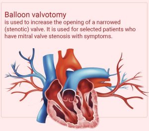
- Balloon Mitral Valvotomy: As mentioned earlier, balloon mitral valvotomy is a minimally invasive procedure in which a balloon-tipped catheter is used to dilate the narrowed mitral valve. It can be an effective treatment option for selected cases of mitral stenosis, especially when the valve anatomy is suitable.
- Mitral Valve Repair or Replacement: In more severe cases of mitral stenosis or when other treatment options are not feasible, surgical intervention may be necessary. Mitral valve repair aims to preserve the patient’s own valve by correcting the abnormalities causing stenosis. In some cases, a mitral valve replacement may be performed, where the diseased valve is replaced with a mechanical or biological prosthetic valve.
- Anticoagulant Therapy: Individuals with mitral stenosis and associated atrial fibrillation or other high-risk factors for blood clot formation may require anticoagulant therapy, such as warfarin, to prevent the formation of blood clots in the heart.
- Antibiotic Prophylaxis: Prior to certain dental or surgical procedures, individuals with mitral stenosis may need to take antibiotics to prevent bacterial endocarditis, which is an infection of the heart lining or valves.
Procedure of balloon mitral valvotomy:
- The patient is typically placed under general anaesthesia to ensure comfort and unconsciousness throughout the procedure.
- A cardiologist inserts a thin, flexible tube called a catheter into a blood vessel, commonly through the groin area. The catheter is guided toward the heart using imaging techniques such as fluoroscopy (real-time X-ray) and echocardiography.
- A guidewire is passed through the catheter and guided across the narrowed mitral valve. The guidewire helps position and guide the balloon to the desired location.
- A deflated balloon, attached to the tip of the catheter, is positioned across the narrowed mitral valve, precisely at the level of the valve orifice.
- Once the balloon is correctly positioned, it is inflated. Inflation occurs by rapidly injecting a sterile liquid, such as contrast dye or saline solution, into the balloon. The inflation pressure exerted by the balloon helps expand the narrowed mitral valve and separate the leaflets, increasing the valve opening.
- Throughout the inflation process, the cardiologist carefully monitors the balloon’s position, pressure, and the effect on the valve using imaging techniques and pressure measurements. This ensures precise and controlled dilation of the valve.
- After an appropriate duration of balloon inflation, the balloon is deflated by removing the liquid from the balloon. Once deflated, the balloon is withdrawn from the heart, along with the catheter and guidewire.
- Following the BMV, the cardiologist evaluates the effectiveness of the procedure by assessing the degree of mitral valve opening and the improvement in blood flow. This assessment is often done using echocardiography.
- After the procedure, the patient is monitored closely in a recovery area or hospital room to ensure stability. Vital signs, heart sounds, and any potential complications are monitored.